Background
The Light Imaging Facility (LIF) provides intramural scientists with access to state-of-the-art light imaging equipment and expertise in light imaging techniques. The Core offers training and assistance in a variety of light microscopic techniques including laser scanning confocal microscopy, video microscopy and digital image analysis. A digital image printer and slide maker are available for preparing illustrations for presentations and publications.
Contact Information
Carolyn Smith, Ph.D. , Core Director
E-mail: smithca@ninds.nih.gov
Scope of Core Facility
The Light Imaging Facility (LIF) is available to all scientists in the NINDS and, as feasible, to other scientists in the Porter Neuroscience Research Center. New users should contact Carolyn Smith to register and for orientation and training.
Using the Light Imaging Facility
- Access: The facility is located in Building 35 (Porter Building). Most of the equipment is on level 2, pods B and C but one of the confocal microscopes is located in the basement (BB520). The LIF also maintains a LSM 510 confocal microscope in Building 49. Facility users will need card key access to Building 35 for entry during evenings and weekends and for entry to Building 49 at any time.
- Register: To register to use the Light Imaging Facility, please contact Carolyn Smith: smithca@ninds.nih.gov
- File storage and transfer: Facility users may store files temporarily on the hard disks of the Facility's computers. A server is available for transferring files between computers, courtesy of the Research Services Branch (RSB). Users are requested to limit their file folders to <100 GB and to delete old files. Be aware that files on the hard disks and server are accessible to others and not secure. Files left on the hard disks or server for longer than six months may be deleted without warning.
- Supplies: The Facility maintains a stock of common reagents and expendables such as fluorescent dyes and secondary antibodies, mounting media, coverslips, and LabTek chamber coverglasses. These are provided free of charge for pilot experiments but users are expected to purchase their own supplies for routine use.
Equipment
Zeiss LSM 800 Confocal Microscope
Location: Porter Building (Bldg. 35) Room 2C401.
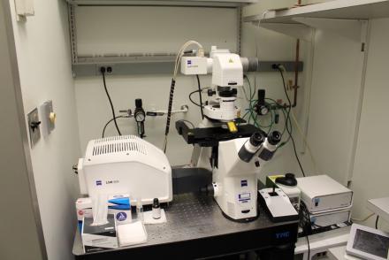
Laser Lines: 405, 488, 561, 633 nm.
Detectors: Two highly sensitive GaASP detectors.
Applications: The Zeiss LSM800 Confocal microscope is well suited for imaging fixed samples such tissue sections. It can acquire images of up to four fluorophores (blue, green, red and far red) by line-by-line wavelength switching or by frame switching. The acquisition software can collect and stitch tiled images of samples too large to fit within the field of view. It can easily be programmed to capture images of multiple regions on a slide or multi-well plate. The high sensitivity of the detector allows detection of weak fluorescent signals while employing low levels of laser illumination and rapid frame rates.
Zeiss LSM 880 Spectral Confocal Microscope
Location: Porter Building (Bldg. 35) Room 2C401.
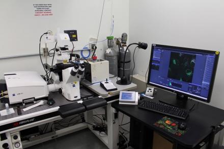
Laser Lines: 405, 488, 561, 594, 633 nm.
Detectors: Thirty-four channel GaASP spectral detector.
Applications: The Zeiss LSM880 Spectral Confocal Microscope is equipped for multi-channel fluorescent imaging in living samples. The spectral detector together with the software for spectral unmixing makes it possible to image simultaneously multiple different fluorophores, provided that reference spectra for each of the individual fluorophores are available. The spectral detector also can be used for conventional multi-channel fluorescence imaging. The high sensitivity of the detector allows detection of weak fluorescent signals while employing low levels of laser illumination. A heating stage and CO2 incubation system are available for maintaining living samples.
About the Spectral Imaging: Spectral_Imaging.pdf
Zeiss LSM 880 AiryScan Confocal Microscope
Location: Porter Building (Bldg. 35), Room 2C403.
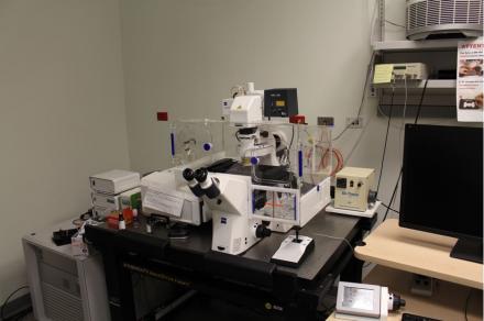
Laser Lines: 405, 488, 561, 633 nm.
Detectors: An AiryScan detector and two standard photomultiplier detectors.
Applications: The Zeiss LSM880 AiryScan Confocal microscope uses a unique 32 channel detector that provides 1.7 fold improved resolution as well as significantly improved signal-to-noise compared to conventional confocal detectors. The high sensitivity of the AiryScan detector makes it possible to detect even very weak fluorescent signals while using low levels of laser illumination. The improved resolution and signal are beneficial for imaging small cellular structures such as synaptic spines, organelles and molecular complexes. However, each color channel must be captured individually and the image acquisition rate is slow. The acquisition time for a stack of three color images from a 3 µm thick sample (16 images with a 1.4 NA objective at 2X zoom) is about 2 min. Acquired images must be processed for optimal resolution in the z-axis.
About the AiryScan Detector: LSM880_Airyscan.pdf
Leica SP8 STED 3X Super-resolution Confocal Microscope
Location: Porter Building (Bldg. 35), 2B810.
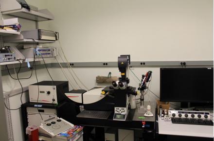
Laser Lines: White Light Laser 470-670 nm. Depletion lasers: 442, 592, 660, 770 nm.
Detectors: Three hybrid detectors, two standard photomultiplier detectors.
Applications: The Leica STED 3X confocal microscope achieves superior resolution compared to conventional confocal microscopes due to the reduced size of the illuminated spot where fluorescence is generated. The gated STED technique reduces the excitation spot to about 50 nm in the horizontal plane and 150 nm in the vertical axis, as compared to 200 nm horizontal and 500 nm vertical for a conventional confocal microscope. Gated STED requires illuminating the sample with two laser beams that together are sufficiently intense to cause considerable photo-bleaching in the sample. Imaging living samples is feasible, but only for a limited number of time points. A newer STED technique based on single photon counting and Fluorescence Lifetime Imaging (FLIM) allows use of lower laser illumination levels and provides superior resolution. The system has both a conventional scanner and a high-speed resonant scanner, useful for imaging rapidly changing specimens, and the "hybrid" detectors provide high sensitivity and dynamic range. The STED technique works well with some but not all conventional fluorophores and fluorescent proteins (see sample preparation protocol to STED Sample Preparation.pdf (attached here(pdf, 6084 KB)).
Suggested fluorophore combinations: DAPI/FITC/Cy3/Cy5; DAPI/Alexa 488/555/647; CFP/YFP; EGFP/DsRed or mCherry. Avoid Alexa 594.
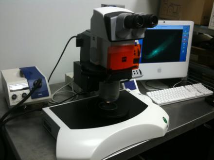
Three Zeiss LSM 510 Confocal Microscopes
Location: Porter Building (Bldg. 35), Room 2B708.
Name/ specifications/ location:
LSM510 405/ three detectors - 405, 458, 488, 514, 543, 633 nm lasers/ Bldg 35, Rm. 2BC909.
LSM510 Meta/ two detectors - 458; 488, 514, 633 nm lasers/ Bldg. 35, Rm 2C109.
LSM510 CF 49/ three detectors - 405, 458, 488, 514, 561, 633 nm lasers/ Building 49, Rm. 3B68.
Applications: The LSM510 confocal microscopes are well suited for imaging fixed samples with blue, green, red and far red fluorophores.
Suggested fluorophore combinations: DAPI/FITC/Cy3/Cy5; DAPI/Alexa 488/555/647; CFP/YFP; EGFP/DsRed or mCherry. Avoid Alexa 594.
GE Typhoon Variable Mode Scanner
Location: Porter Building (Bldg. 35) Room 3A1014.
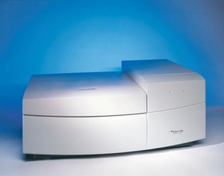
Laser lines: 457, 477, 488, 532, 633
Applications: The Typhoon 9410 Variable Mode Imager is a versatile multi- purpose laser scanner. For DNA, RNA, and protein samples, choose from: storage phosphor autoradiography, direct blue, green or red excited fluorescence, and chemiluminescence. The appropriate optical components are automatically activated when the scanning mode is selected. Typhoon scans wet or dry gels, blots, mounted and unmounted storage phosphor screens up to 35 x 43 cm. It exhibits outstanding linearity, quantitative accuracy, and extremely low limits of detection. Seven user selectable pixel sizes (10, 25, 50, 100, 200, 500 and 1000 microns) ImageQuant TL v7.0 software.
Epsom V850 Flatbed Scanner
Location: Porter Building (Bldg. 35) Room 2B914.
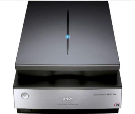
Applications: High resolution scanner for transparent and reflective originals. Connected to an iMac with Adobe Photoshop software.
Imaging Workstations
- Volocity (Perkin Elmer). 3D image visualization, deconvolution and analysis. Measure volumes, track particles, assess colocalization. File formats: tiff, Zeiss lsm and czi, Leica lif, Nikon and others). 35/2B914.
- Metamorph (Molecular Devices). 2D image analysis. Automated morphometry, track particles, create montages and kymographs. File formtas: tiff, Zeiss lsm. 35/2B914.
- Huygens Image Restoration (Scientific Volume Imaging) and Leica Application Suite. Image restoration by deconvolution. Leica 3D image visualization module. File formats: Leica lif, Zeiss lsm and czi, and others). 35/2B814.
- Zeiss Zen Blue and Zen Black. AiryScan image processing, 3D visualization. File formats: Zeiss lsm and czi. 35/2C403.
- Zeiss LSM510 software. Full software package. 35/2B914.
Fluorescence Microscopy Manuals & Tutorials
- Short Guide for LSM510: General procedures for operation of LSM 510(pdf, 2410 KB)
- Volocity User Guide: VolocityuserGuide.pdf(pdf, 9086 KB)
- Metamorph User Guide: Metamorph-User-Guide-Andrea-contents-April-2013.pdf(pdf, 2233 KB)
- Huygens Deconvolution User Guide: HuygensUserGuide.pdf(pdf, 5152 KB)
- Zeiss LSM 800/800 Tutorials: https://www.zeiss.com/microscopy/us/local/zen-knowledge-base/lsm-800-900-980.html
Fluorescence Microscopy Literature
- Basic Confocal Microscopy: An introduction to the principles of laser scanning microscopy and practical tips for confocal users. (pdf, 613 KB)
- Confocal Laser Scanning Microscopy: The theoretical basis of laser scanning confocal microscopy and digital signal processing. (pdf, 961 KB)
- AiryScan Detector: nmeth.f.388.pdf(pdf, 206 KB)
- Super-resolution imaging: Super-Resolution.pdf(pdf, 625 KB)
- Fluorescence microscopy:
- Fluorescence Microscopy, Brian Herman, http://onlinelibrary.wiley.com/doi/10.1002/0471143030.cb0402s13/full
- Principles and Application of Fluorescence Microscopy, Donald Coling, Bechara Kachar, http://onlinelibrary.wiley.com/doi/10.1002/0471142727.mb1410s44/full
Protocols for Sample Preparation
Preparing samples for STED imaging:
Freezing and Freeze substitution for immunofluorescence:
- Spectral imaging: Spectral_Imaging.pdf
- Freeze substitution for immunofluorescence: Freeze substitution for immunofluorescence
Image Analysis Software
Zeiss LSM Image Browser: Image Browser is a free stripped-down version of the LSM 510 software for PC computers. The Zeiss Image Browser can be downloaded from the LIF server (LIF vol 1/Lif vol 1/Zeiss Image Examiner).
ImageJ: ImageJ is a public domain image processing and analysis program developed by Wayne Rasband in the Research Services Branch (RSB) of the National Institutes of Mental Health (NIMH). The program runs on any platform with Java (including Mac OSX) and can be customized through specialized plugin modules or macros. ImageJ can be downloaded from http://rsb.info.nih.gov/ij/. Manual for ImageJ: ImageJ_Manual(1).pdf
Fiji: Fiji is a user-friendly version of ImageJ. It is easy to install, has automatic updates, extensive documentation and lots of plugins for analyzing confocal microscope images and image stacks. Download Fiji: https://imagej.net/Fiji/Downloads
Useful Links
Molecular Expressions: This website is a good source of information about light microscopy, including confocal microscopy. http://www.molecularexpressions.com/primer/index.html
