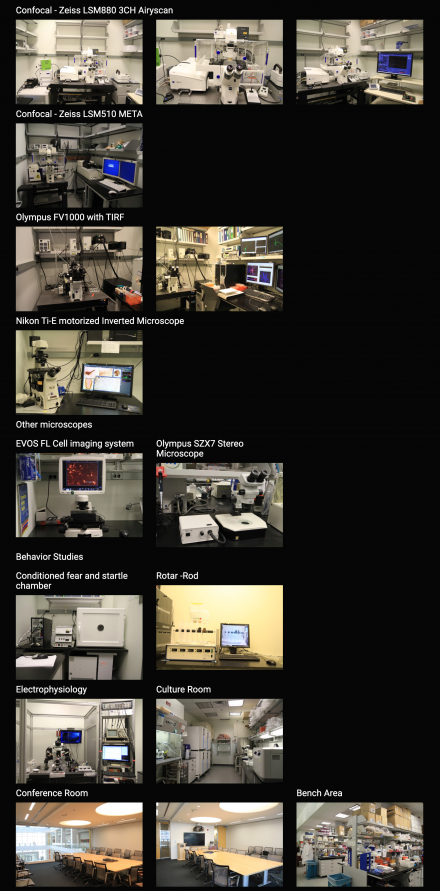
Access to two super-resolution microscopes: Leica SP5 and Leica TCS SP8 STED 3X at NINDS Light Imaging Facility located next door to the lab:
Other NINDS research support facilities:
The PROTEIN/PEPTIDE SEQUENCING FACILITY provides amino acid sequencing of purified proteins/peptides for NINDS investigators. The facility is also available for collaborations involving protein/peptide purification and more complicated sequencing strategies.
The FLOW CYTOMETRY CORE FACILITY provides technical, collaborative and new application research and development support to NINDS and other intramural investigators in both basic and clinical research programs requiring the use of high-throughput conventional and imaging flow cytometry, preparative fluorescence-activated cell sorting and multi-parametric in situ cytometric imaging. The Facility provides high-throughput screening of cells and assaying of their specific biological properties using appropriate biomarkers linked to fluorescent endpoints, in addition to preparative sorting of purified cell populations and/or subpopulations of interest, based on one or more cellular/subcellular parameters, as specified by the investigator.
The LIGHT IMAGING FACILITY provides intramural scientists with access to state-of-the-art light imaging equipment and expertise in light imaging techniques. The Facility offers training and assistance in a variety of light microscopic techniques including laser scanning confocal microscopy, video microscopy and digital image analysis. A digital image printer and slide maker are available for preparing illustrations for presentations and publications.
The EM FACILITY provides intramural NINDS investigators with the opportunity to use electron microscopic techniques in their research programs. The Facility provides assistance in all aspects of electron microscopy including project planning, specimen preparation and training.
