We are primarily interested in the medial entorhinal cortex (MEC), which plays a key role in spatial representation and episodic memory. The MEC is a part of medial temporal lobe and serves as the main gateway between the hippocampal formation and the neocortex. Many functional cell types, including grid cells, head-direction cells, border cells, speed cells, and object vector cells, have been discovered in the MEC and their activity patterns potentially represent spatial and self-motion information during navigation. Dysfunction of the MEC is closely associated with Alzheimer’s Disease (AD), the most common form of dementia. AD patients generally exhibit severe loss of episodic memory and have difficulty in spatial navigation.
Projects in the laboratory are centered around the following questions:
(1) How is spatial information represented and computed in the MEC circuit?
(2) Whether and how is spatial memory encoded in the MEC?
(3) How does the MEC interact with other brain areas to perform its function?
(4) How is the physiological function of the MEC disrupted in Alzheimer’s Disease?
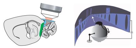
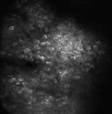
We mostly use mice as model organisms and our experiments integrate optical, behavioral, computational, and molecular approaches. We access the MEC at cellular and sub-cellular resolution using two-photon imaging approach when mice navigate in virtual reality environments (see figures and videos below). This experimental setting provides a great opportunity to measure and manipulate neural dynamics while controlling spatial information during the navigation. In addition, we have a long-standing interest in developing new experimental paradigms to investigate circuit and molecular mechanisms underlying the MEC neural dynamics, as well as probing the function of the MEC in broader cognitive aspects.

Left: we gain cellular-resolution optical access to the MEC (green area) through a microprism, which is surgically inserted into the transverse fissure and reflects the optical path at 90o.
Right: neural dynamics of the MEC is measured when a mouse navigates in virtual reality. The mouse’s motion in a virtual environment is driven by its running on a ball.


